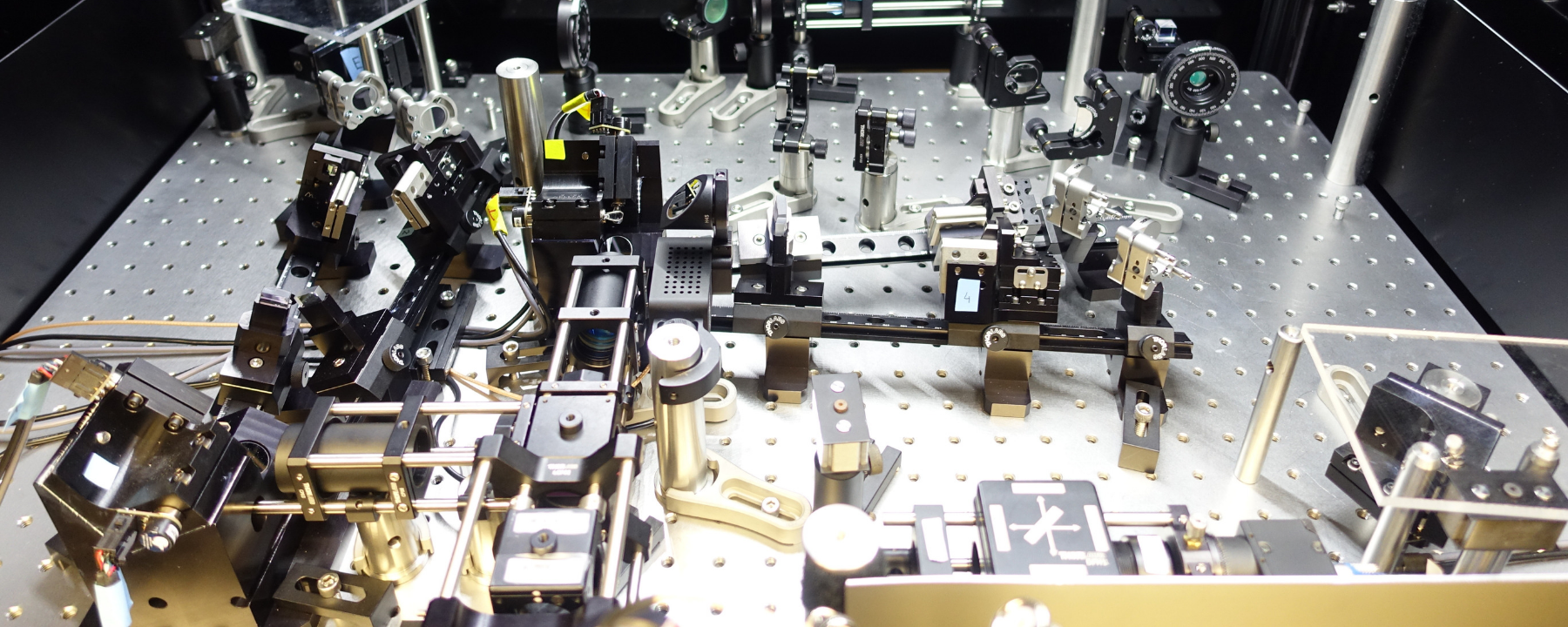
Anew tool called Scanned Line Angular Projection microscopy, or SLAP, enables scientists to record neural activity patterns in the mouse visual cortex, millisecond-by-millisecond, researchers reported July 29 in Nature Methods.
Conventional light microscopy cannot pierce through the dense wrinkles of a living brain, and classic two-photon microscopy takes nanoseconds to process each pixel in a single still image. Creating videos requires researchers to take measurements from every pixel in every frame, and that time adds up quickly. “You’d think that’d be a fundamental [speed] limit—the number of pixels multiplied by the minimum time per pixel. But we’ve broken this limit by compressing the measurements,” says coauthor Kaspar Podgorski, a neuroscientist and cognitive scientist at the Howard Hughes Medical Institute’s Janelia Research Campus, in an announcement.

SLAP works by probing tissue samples with flat planes of light and compressing data from many pixels into a single measurement. Computer algorithms then decompress the data, allowing scientists to render high-resolution videos of brain activity.
A. Kazemipour et al., “Kilohertz frame-rate two-photon tomography,” DOI:10.1038/s41592-019-0493-9, Nature Methods, 2019.
source: here
*
Be the first to comment.